
Sorry, nothing in cart.
Understanding Emergency Medical Treatments
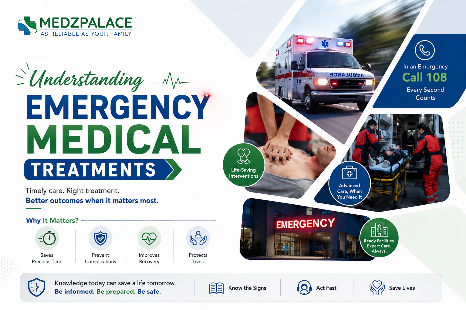
A medical emergency represents a sudden, critical situation that poses an immediate threat to life, limb, or long-term health. In these high-stakes moments, a series of rapid, evidence-based interventions known as emergency medical treatments are deployed to stabilize the patient and prevent catastrophic outcomes. From the actions of first responders at the scene to the coordinated chaos of a hospital emergency department (ED), these treatments form a crucial bridge between a life-threatening event and the potential for recovery. Understanding the principles and applications of these interventions is vital not only for healthcare professionals but also for the public, as swift recognition and action can significantly influence a patient’s prognosis. This article provides a clinical overview of the fundamental concepts and specific treatments that define modern emergency medicine.
The “Golden Hour” and the Chain of Survival
The effectiveness of emergency medical treatments is often dictated by time. Two core concepts underscore this urgency: the “Golden Hour” and the “Chain of Survival.”
The Golden Hour is a term, particularly in trauma care, that refers to the critical window of time following a traumatic injury during which there is the highest likelihood that prompt medical and surgical treatment will prevent death. While not a literal 60 minutes, it emphasizes that a patient’s chances of survival are greatest if they receive definitive care within the shortest possible period. This principle drives the entire pre-hospital and emergency response system, from rapid ambulance dispatch to efficient transport and streamlined ED protocols.
For cardiac arrest, the guiding principle is the Chain of Survival, a sequence of actions that, when performed in rapid succession, maximize the chance of survival. The links in this chain are:
- Immediate Recognition and Activation of the Emergency Response System: Recognizing that a person is unresponsive and not breathing normally, and immediately calling emergency services (e.g., 911, 112).
- Early Cardiopulmonary Resuscitation (CPR): Performing high-quality chest compressions to maintain blood flow to the brain and other vital organs until the heart can be restarted.
- Rapid Defibrillation: Using an Automated External Defibrillator (AED) to deliver an electrical shock to the heart, which can terminate a life-threatening arrhythmia like Ventricular Fibrillation.
- Effective Advanced Life Support: The provision of advanced interventions by paramedics and other healthcare professionals, including advanced airway management and administration of medications.
- Post-Cardiac Arrest Care: Integrated, multidisciplinary care in a hospital setting to treat the underlying cause of the arrest and optimize neurological recovery.
Triage: The First Step in the Emergency Department
Upon arrival at an emergency department, patients do not typically see a physician on a first-come, first-served basis. Instead, they undergo a rapid assessment process called triage. A triage nurse evaluates the patient’s condition to determine the severity and acuity of their illness or injury. This ensures that the most critically ill patients are treated first. Most EDs in the United States use a five-level system, such as the Emergency Severity Index (ESI).
- ESI Level 1 (Resuscitation): Requires immediate, life-saving intervention. Examples include cardiac arrest, severe trauma, or respiratory failure.
- ESI Level 2 (Emergent): A high-risk situation where the patient is disoriented, in severe pain, or whose vital signs are dangerously abnormal. Examples include stroke symptoms, chest pain suggestive of a heart attack, or major fractures.
- ESI Level 3 (Urgent): A condition that requires multiple resources (e.g., labs, imaging) but the patient’s vital signs are stable. Examples include abdominal pain, moderate asthma attacks, or a high fever in an otherwise healthy adult.
- ESI Level 4 (Less Urgent): Requires one resource, such as a simple X-ray or stitches for a minor laceration.
- ESI Level 5 (Non-Urgent): Requires no resources beyond a physician’s examination. Examples include a cold, a minor rash, or a request for a prescription refill.
Core Emergency Medical Interventions
Once a patient is triaged and placed in a treatment room, a team of clinicians begins a more detailed assessment and initiates specific interventions based on the presenting condition.
Cardiopulmonary Resuscitation (CPR) and Advanced Cardiac Life Support (ACLS)
For a patient in cardiac arrest, the goal is to restore a spontaneous heartbeat and circulation. This begins with basic life support (BLS), which includes high-quality CPR—a cycle of chest compressions and rescue breaths. The primary goal of CPR is not to restart the heart, but to manually pump oxygenated blood to the brain to prevent irreversible damage.
Defibrillation is used for “shockable” rhythms like Ventricular Fibrillation (VF) and pulseless Ventricular Tachycardia (VT). An AED or manual defibrillator delivers a controlled electrical current to the heart, depolarizing the entire myocardium and allowing the heart’s natural pacemaker to hopefully resume a normal rhythm.
Advanced Cardiac Life Support (ACLS) builds upon BLS and includes:
- Airway Management: Securing the airway via endotracheal intubation or a supraglottic airway device to ensure effective ventilation.
- Vascular Access: Establishing an intravenous (IV) or intraosseous (IO) line to administer fluids and medications rapidly.
- Pharmacotherapy: Administering drugs such as epinephrine to increase blood pressure and heart contractility, and antiarrhythmics like amiodarone or lidocaine to treat specific abnormal heart rhythms.
- Identifying Reversible Causes: Systematically searching for and treating correctable causes of the arrest, often remembered by the “H’s and T’s” (e.g., Hypovolemia, Hypoxia, Hydrogen ion (acidosis), Hypo/Hyperkalemia; Tamponade, Tension pneumothorax, Toxins, Thrombosis).
Trauma and Hemorrhage Control
In cases of severe trauma, clinicians follow a structured protocol, often known as the Advanced Trauma Life Support (ATLS) primary survey. The mnemonic ABCDE guides the initial assessment and management:
- A – Airway: Assess for airway patency and protect the cervical spine from potential injury.
- B – Breathing: Assess ventilation and oxygenation, identifying and treating life-threatening chest injuries like a tension pneumothorax.
- C – Circulation: Control any external bleeding and assess for signs of shock (e.g., rapid heart rate, low blood pressure). This is the critical step for hemorrhage control.
- D – Disability: Perform a rapid neurological exam, often using the Glasgow Coma Scale (GCS), to assess level of consciousness.
- E – Exposure: Fully undress the patient to perform a complete examination while preventing hypothermia.
The single most important intervention for circulation is stopping major bleeding. Techniques include direct pressure, wound packing with gauze (sometimes impregnated with hemostatic agents), and the application of a tourniquet high on an extremity for catastrophic limb hemorrhage. Internally, a patient in hemorrhagic shock will receive a massive transfusion protocol, which involves the rapid administration of packed red blood cells, fresh frozen plasma, and platelets in a balanced ratio to restore blood volume and clotting ability.
Acute Stroke Management
Time is brain. For every minute an acute ischemic stroke goes untreated, millions of neurons can be lost. Recognizing the signs of a stroke using the BE FAST acronym is a key public health message: Balance loss, Eyesight changes, Face drooping, Arm weakness, Speech difficulty, Time to call emergency services. In the ED, the immediate priority is a non-contrast CT scan of the head to differentiate between an ischemic stroke (caused by a clot) and a hemorrhagic stroke (caused by bleeding). Treatment diverges dramatically based on this finding:
- Ischemic Stroke: If the patient presents within a specific time window (typically 3 to 4.5 hours from symptom onset) and meets strict criteria, they may receive intravenous thrombolysis with a drug like alteplase (tPA), which works to dissolve the clot. For strokes caused by a large vessel occlusion, a procedure called mechanical thrombectomy may be performed, where a catheter is used to physically retrieve the clot from the brain.
- Hemorrhagic Stroke: Treatment focuses on controlling blood pressure, reversing any anticoagulant medications the patient may be on, and consulting a neurosurgeon, as surgical intervention may be required to relieve pressure on the brain.
Myocardial Infarction (Heart Attack) Treatment
When a patient presents with symptoms of a heart attack (e.g., chest pain, shortness of breath, pain radiating to the arm or jaw), the immediate goals are to relieve pain, improve blood flow, and preserve heart muscle. An electrocardiogram (ECG) is performed immediately to look for characteristic changes, particularly ST-segment elevation, which indicates a complete blockage of a coronary artery (a STEMI).
Initial medical treatments often follow the mnemonic MONA, though the order and application depend on the patient’s specific presentation:
- M – Morphine: For pain relief.
- O – Oxygen: If oxygen saturation is low.
- N – Nitroglycerin: To dilate coronary arteries and improve blood flow.
- A – Aspirin: To prevent further clot formation.
The definitive treatment for a STEMI is reperfusion therapy—restoring blood flow to the heart muscle. The gold standard is percutaneous coronary intervention (PCI), where a cardiologist threads a catheter to the blocked artery, inflates a balloon to open it, and often places a stent to keep it open. If PCI is not available within a recommended timeframe (e.g., in a rural hospital), fibrinolytic therapy (“clot-busting” drugs) may be administered.
Management of Severe Allergic Reactions (Anaphylaxis)
Anaphylaxis is a life-threatening, systemic allergic reaction that can cause airway swelling, difficulty breathing, and a precipitous drop in blood pressure (anaphylactic shock). The single most important, first-line treatment is intramuscular epinephrine. Epinephrine works rapidly to constrict blood vessels (increasing blood pressure), relax airway muscles (improving breathing), and reduce the release of further inflammatory mediators. All other treatments, such as antihistamines and corticosteroids, are secondary and do not replace the immediate need for epinephrine.
The Role of Diagnostic Tools in the ER
Emergency treatments are guided by rapid diagnostic evaluations. Key tools include:
- Imaging: Point-of-care ultrasound (POCUS) allows clinicians to quickly visualize organs, detect internal bleeding, or assess cardiac function at the bedside. CT scans provide detailed cross-sectional images essential for diagnosing stroke, pulmonary embolism, and complex trauma. X-rays are used for fractures and diagnosing conditions like pneumonia.
- Laboratory Tests: Blood tests can reveal a wealth of information, from a complete blood count (CBC) to check for infection or anemia, to a metabolic panel to assess electrolytes and kidney function. Cardiac troponin is a specific marker used to diagnose a heart attack.
- Electrocardiogram (ECG): A 12-lead ECG is a non-invasive test that records the heart’s electrical activity and is indispensable for diagnosing heart attacks, arrhythmias, and other cardiac conditions.
Frequently Asked Questions (FAQs)
What is the difference between a paramedic and an EMT?
Emergency Medical Technicians (EMTs) provide basic life support, such as CPR, oxygen administration, and bleeding control. Paramedics have more extensive training and can provide advanced life support, which includes starting IV lines, administering a wide range of medications, and performing advanced airway procedures like intubation.
Why can’t I eat or drink in the emergency room?
You may be asked to refrain from eating or drinking because your condition might require a procedure or surgery that requires an empty stomach to reduce the risk of aspiration during anesthesia. It can also interfere with certain diagnostic tests.
When should I call 911 versus going to an urgent care clinic?
Call 911 or go to the nearest emergency department for any life-threatening or potentially disabling conditions. This includes symptoms like chest pain, difficulty breathing, stroke symptoms (facial droop, arm weakness, slurred speech), severe bleeding, major trauma, or loss of consciousness. Urgent care clinics are better suited for less severe issues like colds, minor cuts, sprains, or urinary tract infections when your primary doctor is unavailable.
Conclusion
Emergency medical treatments are a dynamic and critical component of the healthcare system. They rely on a foundation of rapid assessment, time-sensitive protocols, and a coordinated team approach that spans from pre-hospital care to the emergency department and beyond. While the technologies and medications continue to evolve, the fundamental goal remains unchanged: to intervene decisively at a moment of crisis, preserving life and setting the stage for meaningful recovery.
Medical Disclaimer: The information provided in this article is for educational and informational purposes only and is not intended as medical advice. It is not a substitute for professional medical advice, diagnosis, or treatment. Always seek the advice of your physician or other qualified health provider with any questions you may have regarding a medical condition. Never disregard professional medical advice or delay in seeking it because of something you have read here. In the case of a medical emergency, call 911 or your local emergency number immediately.